History of Present Illness:
A pleasant man in his mid-50’s presents to the hospital after an accident at work where heavy metal containers fell onto his head, chest and both legs. He complains of pain in multiple areas
Vital Signs & Physical Exam:
Vitals stable. Alert. Head, rib, right knee and left ankle pain and tenderness
Initial Diagnostic Testing:
- Imaging: we are focusing on just the right knee for this case
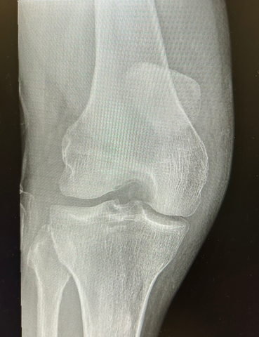
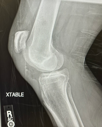
What is the most likely knee injury?
- A) ACL tear
- B) Patellar tendon tear
- C) MCL tear
- D) Tibial plateau fx
- E) Meniscus tear
SCROLL DOWN FOR ANSWERS & 1-MINUTE CONSULT
<<<<<<<<<<<<<<<<<<<<< ADVERTISEMENT & SPACER >>>>>>>>>>>>>>>>>>>>>
****************************************************************************
THE EMERGENCY MEDICINE POCKETBOOK TRIFECTA

- Emergency Medicine 1-Minute Consult, 5th edition
- A-to-Z EM Pharmacopoeia & Antibiotic Guide, NEW 5th edition
- 8-in-1 Emergency Department Quick Reference, 5th edition
***************************************************************************
***************************************************************************
<<<<<<<<<<<<<<<<<<<<<<<<< END SPACER >>>>>>>>>>>>>>>>>>>>>>>>>
ANSWER:
What is the most likely knee injury?
- A) ACL tear
- B) Patellar tendon tear – CORRECT: there is patella alta and some patellar bone fragments
- C) MCL tear
- D) Tibial plateau fx
- E) Meniscus tear
1-Minute Consult on this topic: Click HERE and scroll
CASE CONCLUSION: Also had an ankle fracture on the other side. Admitted for repair of both