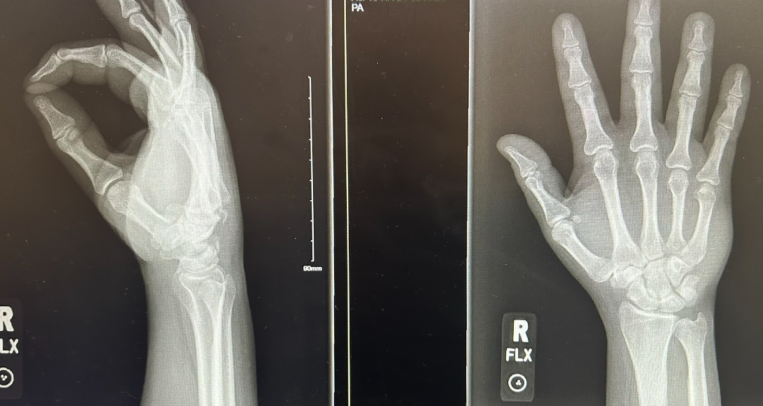
History of Present Illness:

A 52-year-old female with a history of Hep C and cirrhosis presents to the ED in the middle of the Covid pandemic for “coughing up blood”.  She cannot provide a history because is choking and coughing up blood.  Family says she was recently hospitalized for a blood infection.

Vital Signs & Physical Exam:

Bradycardic.  Blood pouring from mouth with each cough

Relevant Test Results:

  • Labs: pending
  • Imaging: CXR

What should you do next

  • A) Intubate, transfuse and call GI
  • B) Intubate, transfuse and call pulmonary
  • C) Keep upright, review imaging from recent admission
  • D) Wait for CBC and CT chest results before intervening

SCROLL DOWN FOR ANSWERS & 1-MINUTE CONSULT

<<<<<<<<<<<<<<<<<<<<< ADVERTISEMENT & SPACER >>>>>>>>>>>>>>>>>>>>>

****************************************************************************

THE EMERGENCY MEDICINE POCKETBOOK TRIFECTA

Emergency Medicine 1-Minute Consult, 5th edition

A-to-Z EM Pharmacopoeia & Antibiotic Guide, NEW 5th edition

8-in-1 Emergency Department Quick Reference, 5th edition 

***************************************************************************

Check Out Our Weekly EM Case Challenges

We’re no longer emailing these but instead are posting on Facebook (ERpocketbooks.com) & Twitter (@EM1MinuteGuru) each Wednesday


***************************************************************************

<<<<<<<<<<<<<<<<<<<<<<<<< END SPACER >>>>>>>>>>>>>>>>>>>>>>>>>

ANSWERS:

  • A) Intubate, transfuse and call GI – Not unless you can prove this is a GI bleed first
  • B) Intubate, transfuse and call pulmonary – Massive hemoptysis patients should not be laid flat and rarely require PRBC’s
  • C) Keep upright, review imaging from recent admission – CORRECT – lying suping can cause asphyxia and cardiac arrest.  You need to try to find out which lung is bleeding and intubate that lung selectively.
  • D) Wait for CBC and CT chest results before intervening – do not send to CT.  She will code when they lay her flat

1-Minute EM Consult on the topic for this case from the Emergency Medicine 1-minute Consult Pocketbook

CLICK HERE TO LEAR MORE ABOUT THIS BOOK

CASE CONCLUSION:  The x-ray image shows bilateral mild infiltrates.  A recent CT showed unilateral cavitary pneumonia from tricuspid valve endocarditis.  She got worse so she had upright selective intubation of her good lung and was taken emergently to the OR for a lobectomy.

CASE LESSONS:

  1. Try to avoid lying hemoptysis patients flat.  Position upright or with bleeding lung down.  If unsure guess and reassess.
  2. Avoid blood and fluids unless patient in obvious hemorrhagic shock.  Shock here was thought to be vasovagal because she was bradycardic.  Here CBC came back normal and the hypotension gradually improved after hemoptysis was treated.  In a case like this keeping the patient a bit hypotensive may minimize further bleeding.
  3. If needs intubation try to Intubate good lung if you can discern which one that is.